Product Description
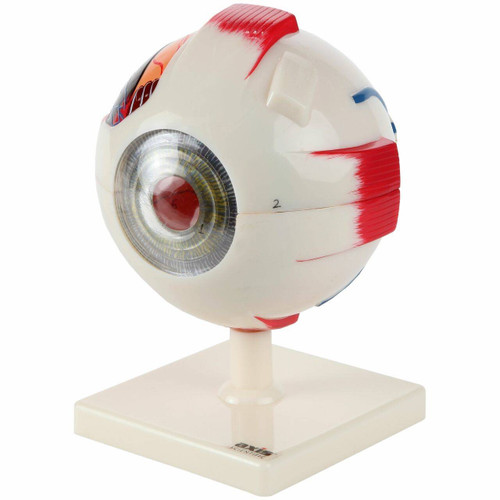
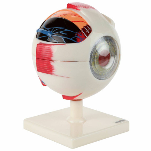
This 5x size , 3-Part Human Ear Model is an enlarged 4D model of the human ear. The ear’s two main functions of balance and hearing are aided by sensory organs and nerves found inside the ear. Common disorders and conditions of the ear include ear infections, tinnitus, and hearing problems. Vertigo is a balance disorder in the internal ear and is a symptom of a more serious condition. This human ear model is enlarged and 4-dimensional, creating a great visualization of the normal anatomy of the ear to aid in diagnostics and treatment of the conditions mentioned.
External Ear
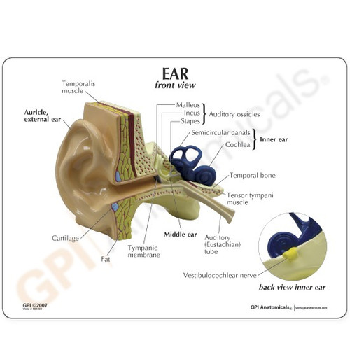
The external or outer ear is represented on the model to help visualize the travel of sound from the external environment to the hearing nerves in the internal ear. Aside from sound, the external ear also provides protection through wax secretion. The enlarged ear model features three parts of the external ear: auricle, external acoustic meatus, and tympanic membrane.
The auricle (pinna) is the visible part of the ear and where sound waves (vibrations) start their travel from the environment and into the ear canal. Featured in the model is the external auditory meatus which extends from the entryway of the ear to the tympanic membrane. The tympanic membrane (eardrum) facilitates the transmission of sound vibrations to the auditory ossicles in the middle ear. Eardrum damage from loud sounds, barotrauma (pressure damage), ear infections, and foreign objects in the ear can result to hearing loss.
Middle Ear
This enlarged ear model features removable auditory ossicles for a detailed study of their anatomy. The model provides a great visualization of how the ossicles fit against the tympanic membrane (eardrum) and the internal ear. This visualization can aid in understanding the physiology and function of the ossicles: transfer vibrations from the eardrum to the internal ear.
As represented by the anatomy model, the middle ear is hollow. This allows for pressure differences outside the body to cause issues. Ear damage (barotrauma) can occur from repeated and drastic changes in pressure. Other disorders of the middle ear include various ear infections, affecting both pediatric and adult patients.
Internal Ear
The 5x enlarged model provides a detailed view of the internal ear and its organs responsible for hearing and sense of balance. The removable internal ear model features the cochlea, semicircular canals, and vestibule. These organs work together with the external and middle ear to maintain balance and transmit sound waves into nerve signals. The removable internal ear features how it fits within the ear and against the other parts.
Internal ear conditions usually affect hearing and balance. Sudden sensorineural hearing loss (SSHL) occurs in the organs of the inner ear or in between the inner ear and the brain. SSHL may happen to one or both ears. Other issues in the internal ear include vertigo, a balance disorder that is an underlying symptom of other conditions.
Base Mount
Our 5x Enlarged 3-Part Human Ear Model is mounted on a sturdy base to fully display a detailed view of the ear’s anatomy.
Full-Color Detailed Study Guide Booklet & User Guide
This Human Ear Model includes a detailed full-color study guide booklet that identifies the 19 anatomical parts featured on the model. All the anatomical parts are numbered on the model and identified in the study guide as an additional in-depth educational resource.
This ear anatomy model features the removable internal ear and auditory ossicles. These removable parts further display specific anatomical features.
Internal Ear:
- Posterior Semicircular Canal
- Anterior Semicircular Canal
- Vestibulocochlear Nerve
- Cochlea
- Oval (Vestibular) Window
- Lateral Semicircular Canal
Auditory Ossicles (Part of Middle Ear):
- Stapes
- Malleus
ustic meatus, and tympanic membrane.
 United States
 Australia
 Canada
 Euro
Euro
 Japan
Japan
 New Zealand
 United Kingdom


